

Tracking Protein Expression Over Time.
- Read More

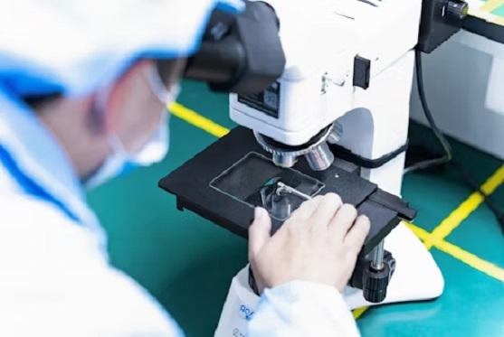
Immunofluorescence microscopy is a powerful technique used to visualize the presence and location of specific proteins or antigens in biological samples using fluorescently labeled antibodies. It's widely used in cell biology, pathology, and diagnostic medicine.
Services:
» Direct Immunofluorescence
» Indirect Immunofluorescence
» Live-Cell Immunofluorescence
» DAPI or Hoechst stains to visualize nuclei
» SYBR® Green or FITC Staining
Immunofluorescence microscopy depicts the full distribution of the epitope that the antibody recognizes and analyzes steady state localization.

These labeled antibodies bind directly or indirectly to cellular antigens. The technique has a number of different biological applications including evaluation of cells in suspension, cultured cells and tissue.
- Infectious Disease Diagnosis
- Autoimmune Diseases
- Cancer Diagnostics and Research
- Genetic Disorders
- Neurological Disorders
The fluorescence can be visualized using fluorescence microscopy. The IF technique allows for a visualization of the presence as well as the distribution of target molecules in a sample. .
Flow Chart of Immunofluorescence microscopy
Sample Preparation (Cell or tissue fixation) > Permeabilizattion > Blocking (To prevent non-specific binding) > Choose Method For Direct or Indirect IFM > Wash again> Mount with antifade solution > View under fluorescence microscope > Analyze & Capture Images > End
Note : Our custom analysis will provide valid solutions and guarantee a high-quality report for your research. Kindly share the technical details of work for quotation and lead time.
Contact: + 91-9891179928| E.mail: info@chemgeneics.org
